Article Topic: The Autonomic Nervous System
Author: Mohammad Khader Altal
Scientific Editor: Marah Bassam Akhdar
Keywords: Autonomic, Sympathetic, Parasympathetic, Enteric Nervous System
Overview and History:
The autonomic nervous system is part of both the central and peripheral nervous systems. It has (afferent, connector, efferent) neurons. The afferent neurons transmit the sensation from the visceral receptor to the CNS where it is transmitted by connector neurons and then the efferent neurons carry the signals to the visceral effector. It is divided into three parts: The sympathetic, parasympathetic, and enteric nervous system(gut brain). Both sympathetic and parasympathetic opposite each other in the effect in most organs and thus they are considered physiological antagonists. However, both systems function in conjunction with one another. It is the balance in the activities that maintain a stable internal environment and homeostasis.
Sympathetic Nervous System:
The sympathetic nervous system is responsible for the “fight or flight” response where the heart rate is increased, arterioles of the skin and intestine are constricted, arterioles of skeletal muscle are dilated, and the blood pressure is elevated. Preganglionic SNS neurons originate from the spinal cord’s thoracic and lumbar regions (T1 to L2) with cell bodies spread around four areas of gray matter, bilaterally and symmetrically, in the spinal cord (1). The preganglionic fiber is short and myelinated while the postganglionic is long unmyelinated. They innervate the eyes and cause mydriasis, as well as the heart, and cause increased heart rate, conductivity, and contractility. They also innervate the GIT smooth muscle and relax them to prevent peristalsis. Additionally, they innervate almost every other organ(2), Figure 1.
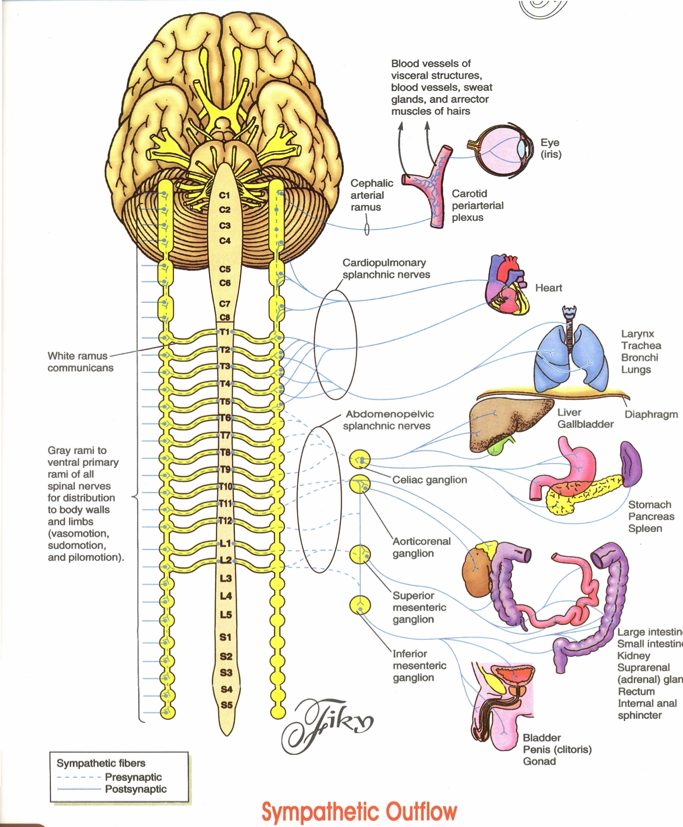
Figure 1: Sympathetic Outflow, (3)
Parasympathetic Nervous System :
The PSNS in contrast to SNS is responsible for the “rest and digest “ state. Also, its preganglionic neurons are long and myelinated while the postganglionic neurons are unmyelinated and short because in the PSNS the autonomic ganglia are very close to the effector organ. The nuclei of the preganglionic neurons are located in the brainstem and the sacral segment of the spinal cord(S2-S4). Moreover, the brainstem nuclei are located in the following cranial nerves: III Oculomotor nerve, VII Facial nerve, IX Glossopharyngeal nerve, X vagus nerve (4), Figure 2. In the “rest and digest” state the PSNS innervates the heart and decreases the heart rate, innervates the GIT and increases peristalsis, innervates the eyes and constricts the pupils, affects the urinary bladder by relaxing the sphincter and constricting its wall, and many other changes (2).
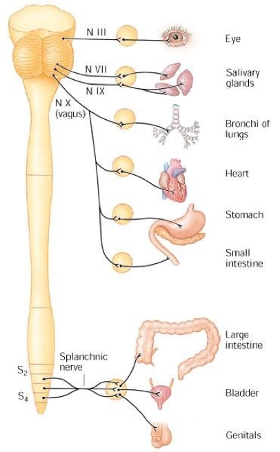
Figure 2: Parasympathetic Nervous System,(5)
Enteric Nervous System:
The enteric nervous system (gut-brain) can function on its own and independently of the SNS and PSNS; however, they do regulate its function. The enteric nervous system is found in the GIT and plays a major role in it. This division is extended along and around the GIT from the esophagus to the anal canal. It is collected in two types of ganglia: The submucosal (Meissner plexus) that lies between the circular muscular layer and the mucosal membrane. This plexus controls glands of the mucous membrane. The second one is the Myenteric (Auerbach) plexus which is found between the circular and the longitudinal muscular layer and controls the muscle movement (Peristalsis) of the gut wall.
Cranial Nerves:
An important part of the peripheral nervous system is the cranial nerves. They emerge from the brain and exit the skull through foramina and fissures to innervate the head and neck. Some of them have either a sensory function or a motor function only while others have both functions. They consist of 12 pairs named as follows:
- Olfactory: It exits through the cribriform plate of the ethmoid bone and has a sensory function(smelling).
- Optic: It exits through the optic canal and has a sensory function(vision).
- Oculomotor: It passes through the superior orbital fissure and has a motor function in the eye muscle such as accommodation, pupil constriction, raising the upper eyelid, and turning the eyeball upward and downward.
- Trochlear: It passes through the superior orbital fissure and has a motor function in the eye by assisting in turning the eyeball downward and laterally.
- Trigeminal: It consists of three branches:*V1(Ophthalmic nerve) passes through the superior orbital fissure and has a sensory function in the scalp, forehead, cornea, etc. *V2(Maxillary nerve) passes through the Foramen rotundum and has a sensory effect in teeth of the upper jaw, etc.*V3(Mandibular nerve) passes through foramen ovale and has both sensory and motor function in muscles of mastication.
- Abducent: It passes through the superior orbital fissure and has a sensory function on the lateral rectus muscle to turn the eyeball laterally.
- Facial: It passes through the internal acoustic meatus, facial canal, stylomastoid foramen and has a sensory function (taste from anterior two-thirds of the tongue), a motor function as well as a parasympathetic function.
- Vestibulocochlear: It passes through internal acoustic meatus and has a sensory function.
- Glossopharyngeal: It passes through the jugular foramen and has a sensory (taste from the posterior third of the tongue and pharynx), motor, and parasympathetic function.
- Vagus: This cranial nerve is special because it innervates the heart, lung, and digestive tract and passes through the jugular foramen. It also possesses significant motor and sensory functions.
- Accessory: It passes through the jugular foramen and has two roots: The spinal roots that innervate the Sternocleidomastoid and trapezius muscles as well as the cranial root the innervates the muscles of soft palate.
- Hypoglossal: It passes through the hypoglossal canal and has a motor function that innervates the tongue and controls its movement and shape (4), Figure 3.
The cranial nerves can be remembered by the following mnemonic:
OOO To Take A Family Vacation Go Vegas After Hour
Cranial Nerves with PSNS Function:
As aforementioned, there are four cranial nerves with PSNS function, and they are:
- Oculomotor: It controls the ciliary muscle (responsible for accommodation) and iris sphincter muscle (for Miosis).
- Facial: It controls the secretion of the salivary glands (sublingual and submandibular) and the glands of the nose and palate as well as the lacrimal glands.
- Glossopharyngeal: It innervates and controls the secretion of the Parotid salivary gland
- Vagus: It innervates the majority of the thoracic and abdominal organs. It decreases the heart rate and increases the peristalsis (smooth muscle contraction) and glandular secretion in GIT.
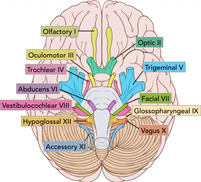
Figure 3:The location of the cranial nerves on the cerebrum and brainstem,(6).
Communicating Rami:
The communicating rami or white communicating branch contains the preganglionic sympathetic nerve and connects it to the spinal nerve. It contains both myelinated and unmyelinated fibers but the myelinated are in higher concentration thus its name (white). They are present on both sides of the spinal column and function ipsilaterally. They originate from the thoracolumbar region (T1-L2) only. In the thoracic region(T1-T12) segment, the thoracic nerve is divided into anterior and ventral rami, and the white rami originate from the anterior rami while in the lumbar region (L1&L2) segment, white rami originate directly from the lumbar nerves. above T1 and Below L2 white rami are absent (7).
Neurotransmitters:
Neurotransmitters are chemical substances that transmit messages from nerve cells across the synaptic cleft to a target cell.
In the autonomic nervous system, the neurotransmitter that is released from the preganglionic fiber in both SNS and PSNS and postganglionic PSNS is Acetylcholine which is quickly terminated(within 2-3 msec) by the hydrolysis action of acetylcholine-esterase.
Ach receptors are found on protein molecules that penetrate the plasma membrane. Once Ach binds to it the structure of the protein will change and an inhibitory or excitatory effect will take place. There are two Ach receptors (cholinergic receptors):
- Nicotinic receptors: They are named so because they respond specifically to nicotine (found in tobacco). These receptors are found predominantly on the preganglionic receptor on both SNS and PSNS and in some postganglionic SNS; such as Sweat glands.
- Muscarinic receptors: They are named so because they respond specifically to muscarine (poison from toadstool fungus). It is found predominantly in the postganglionic receptors of PSNS.
The neurotransmitters that are released from the postganglionic SNS are EN and NE while the neurotransmitter released for the sweat glands is Ach. These receptors are called adrenergic receptors (Alpha1 and 2) and (Beta1-3) (4),(8).
References...