Article topic: Meninges, Ventricular System , and Cerebrospinal Fluid.
Author: Ahmad Aljbali.
keywords: meninges, CSF, choroid plexus ,foramina.
Overview
Meninges:
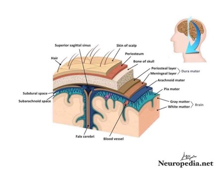
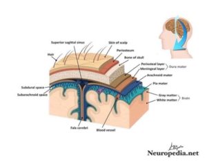
The brain, in the skull (cranial bone), and the spinal cord, in the vertebral column, are enveloped by three protective membranes (meninges): the dura mater, the arachnoid mater, and the pia mater. Based on their location, meninges are referred to as the cranial meninges which surround the brain and
spinal meninges which envelop the spinal cord. Nevertheless, the cranial and spinal meninges are continuous with one another and have the same three meningeal layers. From superficial to deep the
meninges are the:
• Dura mater, also known as the pachymeninx (meninx fibrosa). •Arachnoid mater.
• Pia matter.
These layers bound three clinically important potential spaces:
1.the epidural 2. Subdural 3. subarachnoid spaces.
Meninges role is to protect the brain and spinal cord from mechanical trauma, to support the blood vessels and to form a continuous cavity through which the cerebrospinal fluid (CSF) passes. Specifically, the CSF passes between the inner two meningeal layers (arachnoid and pia) which are together called the leptomeninges.[1]
Meninges of the brain and spinal cord
Meninges of the brain
The dura mater (tough mother)
It is conventionally described as two layers: the endosteal(periosteal origin) layer and the meningeal layer. These are closely united except along certain lines where they separate from each other to form many venous sinuses.
The endosteal layer is the periosteum
covering the inner surface of the skull. At the
foramen magnum, it does not become continuous with
the dura matter of the spinal cord. Recall that, it is around the margins of all the foramina in the skull, it becomes continuous with the
periosteum on the outside of the skull bones. Even at the
sutures, it is continuous with the sutural ligaments .[2][3]
It is most strongly adherent to the bones over the base
of the skull.
The meningeal layer is the dura matter proper. It is a
dense, strong fibrous membrane enveloping the brain and is continuous, through the foramen magnum, with the dura mater of the spinal cord. It provides
tubular sheaths for cranial nerves, as the latter
pass through the foramina in the skull. Outside the skull,
the sheaths of course fuse with the epineurium of the nerves.[2][3]
The meningeal layer sends inward four septa, which
divide the cranial cavity into many freely communicating spaces
that lodge the subdivisions of the brain.[2]
The function of these four septa is to restrict the displacement of
the brain associated with acceleration and deceleration, when the head is moved.
The falx cerebri is a sickle-shaped fold of dura mater that is present in the midline between the two cerebral hemispheres.[2]
Its narrow anterior end is connected to the internal frontal crest and the crista galli. Its broad posterior part blends in the midline with the upper surface of the tentorium cerebelli.
The superior sagittal sinus is in its upper fixed margin, the inferior sagittal
sinus runs in its lower concave free margin, and the straight
sinus runs along its attachment to the tentorium cerebelli.
The tentorium cerebelli is a crescent-shaped fold of
dura mater that is over the posterior cranial fossa. It covers the upper surface of the cerebellum and supports the occipital lobes of the two cerebral hemispheres.
On the anterior side, there is a gap, the tentorial
notch, for the passage of the midbrain, which
produces an inner free border and an outer fixed border. [2]
The fixed border is attached to the posterior
Clinoid processes, the superior borders of the petrous
bones, and margins of grooves for transverse
sinuses on the occipital bone. The free border runs forward
at its two ends, crosses the attached (fixed) border, and is affixed to
the anterior clinoid process on each side. At the point
where the two borders intersect, the 3rd and 4th cranial
nerves pass forward to pass into the lateral wall of the cavernous
sinus (paired Dural venous sinus).[2]
The falx cerebri and the falx cerebelli are attached directly to
the lower and upper surfaces of the tentorium, respectively.
The straight sinus runs down its attachment into the
falx cerebri, superior petrosal sinus walks along its
attachment to the petrous bone, and the transverse
sinus runs along its connection to the occipital bone.
The falx cerebelli is a small, sickle-shaped fold of the dura
mater ,attached to the internal occipital crest, projects forward
between the two cerebellar hemispheres. Its posterior
fixed margin contains the occipital sinus.
Diaphragma sellae is a small, circular fold of the dura
mater that makes the roof of the Sella turcica. A small opening in its center allows passage of the stalk of the hypophysis cerebri .[2]
Arachnoid Mater
The arachnoid mater is a delicate, impermeable membrane,
covering the brain and lying between the pia mater internally
and the dura mater externally. It is separated
from the dura by a potential space, the subdural space,
filled by a film of fluid; it is separated from the pia by the
subarachnoid space, which is filled by cerebrospinal
fluid.
The outer and inner surfaces of the arachnoid are
covered with flattened mesothelial cells.
The arachnoid bridges over the sulci on the surface of
the brain, and in certain situations, the arachnoid and pia
are widely separated to form the subarachnoid cisternae.
The cisterna cerebellomedullaris lies between the inferior
surface of the cerebellum and the roof of the fourth ventricle.
The cisterna interpeduncularis is located between the
two cerebral peduncles. All the cisternae are in free communication
with one another and with the remainder of the subarachnoid space.
In certain areas, the arachnoid projects into the venous
sinuses to form arachnoid villi. The arachnoid villi are most
numerous along the superior sagittal sinus. Aggregations of
arachnoid villi are referred to as arachnoid granulations. Arachnoid villi serve as sites where the cerebrospinal Fluid (CSF) diffuses into the bloodstream.
The arachnoid is connected to the pia mater across the
fluid-filled subarachnoid space by delicate strands of
fibrous tissue.
Structures passing to and from the brain to the skull or
its foramina must pass through the subarachnoid space. All
the cerebral arteries and veins lie in this space, even the
cranial nerve.
The arachnoid fuses with the epineurium of the nerves at their point of exit from the skull.
In the case of the optic nerve, the arachnoid forms a sheath for that nerve which extends into the orbital cavity through the optic canal and fuses with the sclera of the eyeball. Thus, the subarachnoid space extends around the optic nerve as far as the eyeball.[2][3]
The cerebrospinal fluid is produced by the choroid
plexuses within the lateral, third, and fourth ventricles of
the brain. It escapes from the ventricular system of the brain
through the three foramina in the roof of the fourth ventricle
and so enters the subarachnoid space. It now circulates
both upward over the surfaces of the two cerebral hemispheres
and downward around our spinal cord.
Eventually, the fluid enters the blood stream
by passing into the arachnoid villi and diffusing
through their walls.
In addition to removing waste products associated with
neuronal activity, the cerebrospinal fluid provides a fluid
medium in which the brain floats. This mechanism of course
protects the brain from trauma. In addition, the fluid
is now believed to play a role in hormonal transport.[2]
Pia Mater
The pia mater is a vascular membrane covered by flattened
mesothelial cells. It closes to the brain, covering the
gyri and descending into the deepest sulci. It
extends out over the cranial nerves and fuses with their
epineurium. The cerebral arteries entering the substance of
the brain carry a sheath of pia with them.[2][3]
The pia mater forms the tela choroidea of the roof of
the third and fourth ventricles of the brain and it fuses with
the ependyma (thin neuroepithelial) to form the choroid plexuses in the lateral,
third, and fourth ventricles of the brain.[2]
Spinal meninge[1]
Spinal dura mater
Dura mater of spinal cord (Dura mater spinalis) differs from that of the brain by having only one layer; the meningeal layer. The periosteal layer is missing because the vertebral canal, unlike the skull, has its own, true periosteum. The spinal dura mater attaches to the tectorial membrane and posterior longitudinal ligament superiorly. Inferiorly, it extends up to S2 (sacral)vertebral level, thus extending below the spinal cord termination (L1/L2).

The space between the spinal dura mater and the periosteum of the vertebral column is called the epidural space. It is filled with loose connective and adipose tissues, and traversed by the anterior and posterior internal vertebral venous plexuses.
Spinal arachnoid mater
Arachnoid mater of spinal cord (Arachnoidea mater spinalis) is continuous with that of the brain. It lies close and beneath the spinal dura with a narrow subdural space existing between them. Deep to the arachnoid, is the spinal pia mater. Between arachnoid and pia, is the spinal subarachnoid space. This space expands at the level of the conus medullaris (conus terminalis) of the spinal cord, forming the lumbar cistern.
The lumbar cistern extends from L1-S2 and it contains the dorsal and ventral rootlets of some spinal nerves (cauda equina). It is clinically significant as it is the site of lumbar puncture (extraction of CSF for biochemical, microbiological and cytological analyses).
Spinal pia mater
Pia mater of spinal cord (Pia mater spinalis) continues onto the crani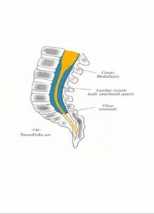 al pia exactly at the level of the foramen magnum. It closely envelops the spinal cord, containing a vascular plexus for the spinal cord tissue. From the apex of the conus medullaris, the pia mater gives off a fibrous projection called the filum terminale (terminal thread). The filum terminale extends around 20 centimeters downwards and attaches to the periosteum of the first coccygeal vertebra.[1]
Starting from the level of the foramen magnum to the level of vertebra T12, the spinal pia shows 21 pairs of ligamentous lateral projections that pass through the arachnoid and attach to the spinal dura mater. These projections are called the denticulate ligaments. Each pair of denticulate ligaments (triangular shaped ligaments) is located halfway between the successive pairs of the spinal nerves. The function of denticulate ligaments is to position and hold the spinal cord in place.[1]
Meningeal spaces
The meningeal spaces are the spaces between the meningeal layers. There are three clinically significant meningeal spaces; epidural (potential), subdural (potential), and subarachnoid (significant one with CSF).
Epidural space
Epidural space (Spatium epidural)
Remember that “Epi” is a prefix indicating that something is “above”. Thus, it should come easy to remember that the cranial epidural space is a potential space between the superficial layer of dura mater and the calvarium (skull). On the other hand, the spinal epidural space is located between the spinal dura mater and the tissues that line the vertebral canal.
The spinal epidural space is a site of applying the local epidural anesthesia. The procedure may be performed at any vertebral level, and the choice depends on the body region that is desired to be anesthetized for any upcoming surgical/obstetric procedure. The applied anesthetics anesthetize locally the spinal nerve rootlets resulting in analgesia (pain relief).[1]
Subdural space
Remember that “Sub” is a prefix that tells us that something is “below”. So, the subdural space is a potential space between the dura mater and the underlying arachnoid mater. The spinal subdural space is continuous with the cranial subdural space. They’re both very narrow and likely contain a thin film of fluid.[1]
Subarachnoid space
Subarachnoid space (Spatium subarachnoideum) is a space between the arachnoid and pia matter. The subarachnoid space contains cerebrospinal fluid (CSF) and major blood vessels and provides expansions known as cisterns. The subarachnoid spaces of the cranium and vertebral column are continuous with each other, creating a closed route for the cerebrospinal fluid(CSF) circulation. Let’s review the route of the cerebrospinal fluid in order to understand the continuity of the subarachnoid space.[1]
Ventricular System and Cerebrospinal Fluid
The CSF is formed by the cells of the choroid plexus within the walls of the brain ventricles. The fluid passes from the two lateral to the third ventricle, and then directly to the fourth ventricle.
From the fourth ventricle, the CSF passes into the central canal of the spinal cord and into the interpeduncular and quadrigeminal subarachnoid cisterns.
The CSF then reaches the subarachnoid space of the brain and spinal cord, circulating through them.
Finally, the CSF is reabsorbed into several dural venous sinuses by diffusing through the subarachnoid granulations in the cranial subarachnoid space.
So after this brief we discuss some details :
Ventricular system
The ventricular system in the brain is composed of (CSF)-filled ventricles and their connecting foramina.[4]
• lined with ependyma and contain CSF.
• contain choroid plexus, which produces CSF at a rate of 500 to 700 ml/day.
• communicate with the subarachnoid space via three foramina in the fourth ventricle.
• consist of four fluid-filled communicating cavities within the brain.
.There are several foramina, openings acting as channels, that connect the ventricles to each  other.
figure(1-4) foramina of ventricular system .[5][6]
A. Lateral ventricles
• the two ventricles are located within the cerebral hemispheres.
• communicate with the third ventricle via the interventricular foramina.
• consist of five parts:
1.Frontal (anterior horn)
• located in the frontal lobe; its lateral wall is formed by the head of the caudate nucleus.
• lacks choroid plexus (no CSF production).
2.Body
• located in the medial portion of the two lobes(the frontal and parietal lobes).
• contains choroid plexus.
• communicates with the third ventricle via the interventricular foramina as in the above figure.
3. Temporal (inferior) horn
• located in the medial part of the temporal lobe.
• contains choroid plexus.
4.Occipital (posterior) horn
• located in the parietal and occipital lobes.
• lacks choroid plexus.
5. Trigona (atrium)
• found at the junction of the body, occipital horn, and temporal hor nof the lateral ventricle.
• contains the glomus (a large tuft of choroid plexus) which is calcified in adults and is visible on x-ray film and computed tomography (CT).
B. Third ventricle
• a slit-like vertical midline cavity of the diencephalon.
• communicates with the lateral ventricles via the interventricular foramina and with the fourth ventricle via the cerebral aqueduct.
• contains choroid plexus in its roof.
C. Cerebral aqueduct (aqueduct of Sylvius)
• lies in the midbrain.
• connects the third ventricle with the fourth ventricle.
• lacks choroid plexus.
• Blockage leads to hydrocephalus (aqueductal stenosis) that means an accumulation of cerebrospinal fluid (CSF) occurs within the brain.
D. Fourth ventricle
• lies between the cerebellum and the brainstem.
• contains choroid plexus in the caudal aspect of its roof.
• expresses CSF into the subarachnoid space via the two lateral foramina and the single medial foramen.
Cerebrospinal Fluid
• a clear, colorless, acellular fluid found in the subarachnoid space and ventricles.
A. Function
• supports and cushions the central nervous system (CNS) against concussive injury (shock absorber).
• transports hormones and hormone-releasing factors.
• removes metabolic waste products through absorption; the sites of greatest absorption are the arachnoid villi (especially, when CSF pressure is elevated).[6]
B. Composition
• contains not more than 5 lymphocytes/microliter and is usually sterile.
• other normal values are:
1. pH:7.35
2. Specific gravity: 1.007
3. Glucose: 66% of plasma glucose
4. Total protein: <45 mg/dl in the lumbar cisterna.[6]
C. Formation
• produced by the choroid plexus at a rate of 500 to 700 ml/day. The total CSF volume is approximately 140 ml .[6]
D. Circulation
• flows from the ventricles via the three foramina of the fourth ventricle into the subarachnoid space and over the convexity of the cerebral hemisphere to the superior sagittal sinus, where it enters the venous circulation.
E. Normal pressure
• is 80 to 180 mm of water (CSF) in the lumbar cistern when the patient is lateral recumbent position.[6]


