Neurosurgery Insights

Dr. Omar Jbarah
Conducted by:
Dr. Omar Jbarah
Neurosurgery Specialist, Founder and General Director of Neuropedia, Fellow in Cerebrovascular Neurosurgery (Japan)
Session 1: Introduction to Neurosurgery & Its Importance
- Overview of Neurosurgery:
- Definition and scope (brain, spine, peripheral nerve surgeries).
- Why neurosurgery is critical for patient care.
- Role of a Neurosurgeon:
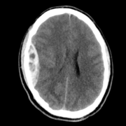
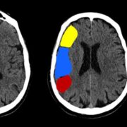
- Lifesaving interventions: brain trauma, tumors, vascular conditions.
- Examples of high-impact neurosurgical cases.
- Neurosurgical Subspecialties:
- Cranial surgery, spine surgery, pediatric neurosurgery, functional neurosurgery , etc.
- Interactive Q&A: Why attendees are interested in neurosurgery.
Session 2: Hot Topics in Neurosurgery
- Brain Surgery:
- Vascular Neurosurgery ( Aneurysm , AVM , etc. )
- Brain Tumor Surgery (e.g., Gliomas, Meningiomas).
- Minimally Invasive Techniques (Endoscopic Surgery).
- Functional Neurosurgery: Deep Brain Stimulation (DBS) for Parkinson’s, epilepsy surgery..
- Spine Surgery:
- Degenerative Spine Conditions (herniated discs, stenosis).
- Minimally Invasive Spine Surgery.
- Real-life Cases: Patient journey from diagnosis to recovery.
- Discussion: How technology is shaping the future of neurosurgery.
Session 3: Pathway to Becoming a Neurosurgeon
- The Training Journey:
- Medical School Requirements.
- Neurosurgery Residency (years, structure, challenges).
- Subspecialty Fellowships (cranial, spine, pediatric, etc.).
- Essential Skills: Technical proficiency, problem-solving, teamwork.
- Daily Life of a Neurosurgeon:
- Case examples: Trauma care, emergency surgeries, elective surgeries.
- Stress and rewards: The high stakes and emotional fulfillment of the job.
- Why We Need Neurosurgeons:
- Addressing the global shortage.
- Neurosurgeons as pioneers of innovative treatments.
- Q&A: Navigating the path and career motivations.
Session 4: Life After Becoming a Neurosurgeon
- Career Paths:
- Clinical Practice vs. Research vs. Academia.
- Global Health and Neurosurgery: Humanitarian missions, making a difference in underserved areas.
- Challenges and Rewards:
- Balancing personal life and career.
- The emotional resilience needed for high-pressure situations.
- Opportunities for continuous learning and advancement in neurosurgery.
- Future of Neurosurgery:
- What’s next? AI, VR, and cutting-edge advancements.
- Telemedicine and global collaboration.
- Panel Discussion with Neurosurgeons:
- Insights from established neurosurgeons on their journey and advice for students.
Course Structure:
- Start Date : 4-10-2024 (Friday)
- Duration: 1.5 – 2 hours weekly sessions (Every Friday – 6:00 PM )
- Format: Online Sessions – 30-minute presentation followed by 60 minutes of interactive discussion, Q&A, and case studies.
- Goal: To provide students with a clear understanding of neurosurgery as a career, its innovations, and the vital role it plays in healthcare.
- Fees: 50 JD – Cliq or Zain Cash Transfer to 0795069358