Article topic: Autonomic Nervous System
Author: Ayah Bani Mustafa
Editor: Yazan S. Al-Zamer, Ethar Hazaimeh.
Keywords: Autonomic, Sympathetic, Parasympathetic, Enteric Nervous System
Overview
The nervous system is divided into the central nervous system (CNS) and peripheral nervous system; the latter is also divided into the afferent and efferent nervous system, the autonomic nervous system (ANS) is a part of the efferent system. The autonomic nervous system transmits electrical impulses through nerve fibers that terminate at effector cells (most body organs), exerting a variety of effects specific to different organs. The autonomic outputs may be divided into the sympathetic, parasympathetic, or enteric systems.
ANS regulates many everyday requirements of vital body functions (including heart rate, blood pressure, respiration, digestion, gland secretions, urination, and sexual arousal) without consciousness, Because of the involuntary nature of the ANS and its functions, it is also known as the visceral, vegetative, or involuntary nervous system.
Anatomy and position of ganglia and fibers :
The sympathetic nervous system (SNS) and the parasympathetic nervous system (PNS) contain both are composed of:
- Afferent neurons: which transmit sensory input by cranial nerves toward the CNS (hypothalamus/brainstem and spinal cord mainly ). this input is analyzed and a response is made out by the transmission of nerve signals that influence the activity of efferent neurons ( specifically preganglionic nerve) of the system. (1) It is an important part of the reflex regulation of the ANS.
- Efferent neurons: which is the motor output of the system, the autonomic nervous system transmits its nerve impulses from the central nervous system to the effector organs by two means: the preganglionic neuron’s fibers and the postganglionic neuron’s fibers. The cell body of the preganglionic neuron is in the CNS, emerging out from either the brainstem/spinal cord (specifically from the lateral horn of the gray matter ) and it makes a synapse in ganglia (which is an aggregation of a large number of nerve cells found outside the central nervous system), its functions include the transmission of signals from the preganglionic neurons to the postganglionic neurons. The postganglionic neuron, leaving out from the ganglia, innervates the effector tissue, It is a nonmyelinated neuron and terminates in effector tissue making a neuro-effector junction, where the axons of these nerve fibers enter a specific tissue, they end up in a number of swellings known as varicosities. When neurons are stimulated by any means, these varicosities release specific neurotransmitters along the whole length of the axon and over a large area in the target organ. The neurotransmitter diffuses and binds to its specific receptors that are located in the target tissues. This diffused type of release of neurotransmitters affects many tissue cells at the same time. (1)(2) preganglionic neurons of both divisions of the ANS (the SNS and PNS) release acetylcholine (ACh) as their neurotransmitter. Postganglionic sympathetic neurons mainly release norepinephrine (NE) as their effector neurotransmitter to affect target organs or they could release epinephrine, while postganglionic parasympathetic neurons release Acetylcholine as their only neurotransmitter. (2)
The efferent ANS is also divided:
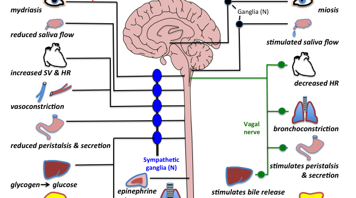
1. Sympathetic neurons: The sympathetic and parasympathetic neurons both emerge from the CNS and start from two different spinal cord origins. The preganglionic neurons of the sympathetic system origins extend all the way from the thoracic and lumbar regions (T1 to L2) of the spinal cord, and they make a junction in two chains of ganglia, preganglionic neurons of the sympathetic system are highly branched allowing each preganglionic neuron to interact and connect with multiple postganglionic neurons, which usually functions as one unit. The preganglionic neurons are short in comparison to the postganglionic ones. neurons of the postganglionic neuron extend from these ganglia to the target tissues that they innervate. The adrenal medulla (considered ganglia–like) receives preganglionic neuron fibers and responds to stimulation by releasing epinephrine (mostly ) or norepinephrine directly into the blood. The sympathetic nervous system mediates a ‘ fight or flight reaction. Postganglionic sympathetic neurons release Norepinephrine that acts on adrenergic receptors in the target tissue. The subtype of the receptor, alpha-1, alpha-2, beta-1, beta-2, or beta-3. The effect is to increase heart rate and blood pressure, to mobilize energy stores of the body to produce free energy, and to increase blood flow to skeletal muscles and the heart while reducing blood flow to the skin and internal organs. Sympathetic stimulation mediates the dilation of the pupils and the bronchioles. It also reduces GI motility and the function of the bladder and sexual organs(2). It also increases renin secretion from the kidney leading to increased intravascular volume(3)
2. Parasympathetic neurons: The parasympathetic preganglionic fibers originate from cranial nerves III, VII, IX, and X and from the sacral region (S2 to S4) of the spinal cord and junction in ganglia near/on the effector organs. Thus, in contrast to the sympathetic neurons, The preganglionic neurons are longer in comparison to the postganglionic ones. Since the preganglionic nerve is not branched, It is able to make a one-to-one connection between preganglionic and postganglionic neurons, allowing a more discrete response for every neuron. The parasympathetic nervous system mediates a ‘rest or digest ‘reaction. This is required for maintaining life, since it maintains essential bodily functions, such as digestion and elimination of wastes, promotes cardiac relaxation, promote salivation, contraction of eye muscles, constriction, and increased secretion of trachea and bronchioles, and increased motility of the gastrointestinal tract. Postganglionic parasympathetic neurons release Acetylcholine that acts on muscarinic and nicotinic receptors, each with various subunits: M1, M2, M3, and N1, N2(for sweat glands innervations). (2)
3. Enteric neurons: It is an intrinsic collection of nerve fibers that entirely innervate the gastrointestinal (GI) tract, plus the pancreas, and gallbladder, also known as the “brain of the gut.” It is made of two plexuses 1) the myenteric (Auerbach) plexuse and 2) the submucosal (Meissner) plexuse.(2) The enteric nervous system all as a whole originate from neural crest cells (4). This system works independently of the CNS and regulates the functions of motility, exocrine and endocrine secretions, and microcirculation of the GI tract. It is regulated by both the sympathetic and parasympathetic nervous systems.
Most body organs are innervated by both the sympathetic and parasympathetic systems. Usually, one system effect is dominating over the other. This organization is finely tuned to regulate the homeostatic function of the body. The exceptions are the adrenal medulla, kidney, sweat gland, poliomotor muscle, which are innervated only by the sympathetic nervous system.