Neurology

Dravet syndrome

5
(4)

Title of article: Dravet syndrome
Authors: Hossam Salameh, Yasmeen Dahabreh
Organizers: Ainaa A.Alzamari, Hamid Ghanem
Reviewer: Ethar Hazaimeh


Keywords: Dravet syndrome(DS), severe myoclonic epilepsy of infancy (SMEI), SCN1A, NAV1.1, Febrile seizures, sudden unexpected death in epilepsy (SUDEP), genetic epilepsy with febrile seizures plus (GEFS+), status epilepticus (SE), multiple anti-seizure medications (ASMs).                


Abstract

Background: Dravet syndrome (DS) is a genetic refractory epilepsy, formerly known as severe myoclonic epilepsy of infancy. It is characterized by temperature-sensitive seizures, resistance to therapy, and developmental differences in children. Despite meeting developmental milestones before seizures, they show signs of autism spectrum features and developmental delays. Ninety percent of children with Dravet syndrome have a pathogenic mutation in the SCN1A gene, impacting the function of certain regulatory neurons.

 Methods: A systematic review search was done which studied Dravet syndrome(DS) primarily through the PubMed database.

 Conclusion: Dravet syndrome is a rare type of epilepsy that usually starts in the first year of a child’s life. In addition to frequent and varied seizures, Its first seizure has a long time occurrence and is triggered by a high fever in most cases. About 90% of DS children have a de novo SCN1A mutation while in 10% of the cases, the variant is inherited

  Introduction

Dravet syndrome (DS) also known as Severe Myoclonic Epilepsy of Infants (SMEI) (1) is a genetic refractory or intractable epileptic syndrome. More than 80% of cases of DS have the SCN1A gene mutation (2,3), which codes for the alpha-1 subunit in the voltage-gated sodium channels (NaV1.1) mainly affecting inhibitory GABAergic interneurons causing dysfunctional channels, which function to regulate the firing in the brain circuits and therefore are essential for the inhibitory regulation, ultimately leading to exaggerated firing with little opposing regulatory impulses (4,5). DS usually initially manifests as prolonged febrile and afebrile seizures during the first year of life. During the second year, it can be understandable that developmental impairments would emerge (6) as the patient would show signs of developmental issues at that age. Premature death occurs approximately in 10-20% of the cases as a consequence of status epilepticus (SE), sudden unexpected death in epilepsy (SUDEP), and acute encephalopathy (4).

Pathophysiology

Dravet syndrome (DS) is primarily caused by SCN1A loss of function mutations, which were identified in 2001(7). The SCN1A gene codes for the voltage-gated sodium channel alpha subunit (Nav1.1).Animal studies have shown that NaV1.1 expression mainly occurs in inhibitory interneurons, which control the excitability of pyramidal neurons. The loss of Nav1.1’s function minimizes the activity and firing of GABAergic inhibitory neurons, leading to hyperexcitability in the brain(8). Low levels of functional NAV1.1 can decrease inhibitory circuitry controlled by parvalbumin-positive interneurons(PV), potentially causing seizures. Inactivation of this protein can also decrease seizure threshold, making it more likely to cause seizures(9). 

Although the etiology of DS resulting from SCN1A mutations is not fully understood, the “inhibitory interneuron hypothesis” and dysfunction of inhibitory interneurons remain the most widely recognized theory. The fundamental cause of DS epilepsy is thought to be haploinsufficiency of Nav1.1 due to pathogenic mutations in SCN1A, which results in the malfunctioning of inhibitory interneurons(10,11). Additionally, loss of NaV1.1 function in cerebellar Purkinje neurons may contribute to ataxia, a common comorbidity of DS(12).

 About 80% of Dravet syndrome (DS) patients have the SCN1A variant, which is a genetic condition affecting over 1000 patients(13). Over 50% of patients have nonsense, frameshift, and splice sense variants, while missense mutations occur in one-third to one-half of cases(14,15). Some patients are negative for SCN1A anomalies on sequencing and have rare duplications and amplifications affecting the gene(16). 90% of DS cases are caused by de novo SCN1A mutation, with 5-10% being of familial inheritance due to parental mosaicism(17). However, the correlation between genotype and phenotype is weak(18).

The type or location of the SCN1A mutation may impact the severity of clinical manifestations. Missense mutations are linked to a less severe phenotype and are negatively correlated with a severe phenotype than nonsense mutations(19). Individuals with mosaic truncating (nonsense) variants are less severely impacted than those with heterozygous truncating variants(20). Familial inheritance is negatively connected with a severe phenotype(21). More severe phenotypes are linked to mutations in the pore region(21,15). Truncating mutations are linked to an earlier age at which myoclonic seizures begin, atypical absence seizures, and a higher rate of cognitive deterioration(14,15). Nonsense variants or variants that impact the sodium channel’s pore-forming area are closely associated with gait deterioration, specifically crouched gait(22).

 Clinical presentation

Dravet syndrome (DS) is a rare epilepsy disorder characterized by refractory epilepsy, neurodevelopmental delay, and cognitive and motor dysfunction(23). The majority of DS patients have seizure-triggering factors, with the most common causes being factors leading to hyperthermia, such as physical exercise or fever(24). The initial seizure accompanied by fever is considered characteristic(4), but only 55% of DS patients experienced fever at their first seizure onset(25). DS patients often experience recurrent febrile or afebrile seizures in the months following the initial events, which can delay diagnosis(25,26). 

DS is classified as a combined generalized and focal epilepsy [27], as both generalized and focal seizures occur. The initial seizure usually occurs in the latter half of the first year, typically between the fifth and eighth month of life. The first seizure is usually bilateral tonic-clonic, a motor seizure that begins in a focal area in the brain and involves both sides [28,29]. It may develop into status epilepticus (SE), a state of inability to terminate an ongoing seizure [30]. It is rare for the initial seizure to be myoclonic or focal, characterized by irregular arrhythmic jerky movements, which might not be acknowledged as epileptic and precede the first convulsive seizure in days or weeks [28,31].

Despite anti-seizure medication, recurring episodes of seizures may still occur in the weeks and months after the initial event [32], with psychomotor impairments first noticeable months after the onset. In the first five years of life, DS patients may experience various types of seizures due to a decreased threshold triggered by internal or external factors [23]. These seizures include myoclonic, absence, obtundation status, focal, and tonic seizures. Neurologic signs such as hypotonia, ataxia, pyramidal signs, dysautonomic signs, and loss of regulation through sweating begin to appear, with common findings being hypotonia, ataxia, pyramidal signs, and dysautonomic signs [33-36].

DS is a condition where patients experience developmental delay, which typically begins after the first seizure and becomes noticeable after the second year of life [37]. DS patients have prolonged developmental progression, with regression or loss of learned skills detected[38]. They typically reach a level in language development where they can speak but fail to construct simple sentences. Cognitive immaturity is evident in the first five to six years of life but not all patients have the same level of impairment [39,40].

Behavior disturbances in individuals with Dravet syndrome (DS) often occur in childhood, causing hyperactivity, attention deficit, and underdeveloped social skills. Motor and cognitive impairments also impact social life [39,28,41]]. In later life, most DS patients experience seizures and varying cognitive disabilities, with some having less severe forms and better functioning [22,42,43]. Seizures and episodes of SE in adulthood tend to decrease in severity and frequency, with convulsive seizures mainly occurring during sleep and focal, myoclonic, and atypical absence seizures potentially disappearing in the future [44].

EEG findings in older patients are normal, but severe motor deterioration can lead to increased theta frequency activity, particularly in central regions and vertex. Motor dysfunction can range from mild to severe, including ataxia, tremors, improper fine movements, dysarthria, spasticity, hyperreflexia, and parkinsonian signs [22,43,45-47]. Gait impairments are also present in 50% of drowsy patients, with a “crouched” gait pattern and wide-based gait. Postural abnormalities and gait disturbances worsen with age, and patients may develop skeletal abnormalities such as flat feet, claw feet, kyphosis, and kyphoscoliosis [47,48-50].

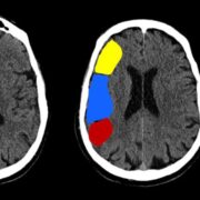
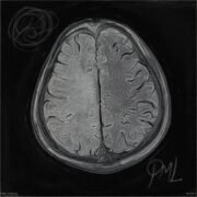
 DS patients’ mental health improves with behavioral disturbances, but many go undiagnosed and untreated [38,47]. DS can cause mortality at any age, especially at younger ages due to SE or SUDEP episodes. Cardiac function in DS patients can differ from controls or those given anti-seizure medication. Brain imaging remains normal, but with age, abnormalities like hippocampal sclerosis, cerebrum and cerebellum atrophy, cortical dysplasia, and increased white matter signaling may occur[34,45,51,52].

 Diagnosis

To clinically diagnose a DS patient, as mentioned before, a certain pattern of signs and symptoms would suggest such a diagnosis, and these are being previously healthy, having recurring drug-resistant seizures [23,53,54], with the typical setting of high-grade fever triggering such events early in life, associated with behavioral and intellectual disabilities that become evident later on, neurological and motor disabilities often manifest as hypotonia and crouch gait along with improper dexterity, and developmental regress and delay especially in the following weeks after a lengthy seizure episode [23,55]. 

To confirm a case of DS, genetic testing is a must and is considered confirmatory and recommended in situations where clinical features are not sufficiently indicative [56]. Neuroimaging techniques using EEG and MRI are used in suspected cases of DS [55], however, EEG and clinical diagnosis based on the above-mentioned clinical features are sufficient to diagnose DS clinically [55,57].

 Gene testing is a cost-effective method for diagnosing epilepsy, as it can help avoid misdiagnosis due to similarities with other syndromes. A negative test result is not enough to exclude epilepsy, especially if clinical characteristics are evident. Early confirmation of a genetic involvement is crucial, especially for patients with sodium channel-blocking anti-seizure medication. However, confirming an SCN1A variant is not enough to confirm epilepsy, as it could be associated with other conditions. Using a gene testing panel for epilepsy is essential for accurate diagnosis(23,58)

Differential diagnosis

Differential diagnosis of DS varies depending on the disease phase, with diagnosing DS after a few febrile seizures being challenging(59). Adult patients may no longer exhibit typical myoclonus and myoclonic seizures, especially if medical records are not available(59). DS can be diagnosed using various tools, including EEG, neuroimaging, and a developmental history of seizures(59).

Differentiating between DS first clonic seizures and febrile seizures or genetic epilepsy with febrile seizures plus (GEFS+) is crucial, as DS seizures are often associated with fever. DS onset is early before completing the first year of life, and the seizure type is clonic and often unilateral whereas it is generalized in febrile seizures, where the duration of the seizures is prolonged and may evolve into status epilepticus and the temperature needed to induce the seizure is not very high(60).

Patients who have epilepsy with myoclonic–astatic seizures (Doose syndrome) are distinguished by having various seizure types like generalized tonic-clonic seizures, myoclonic,myoclonic-atonic, and frequent “drop attacks” that start between the 6th month to the 6th years (typically begins after the age of two). Drop-attacks are uncommon in Dravet syndrome and have a different course(61). Atypical absence status with myoclonic jerks occurs, but there are no focal seizures nor focal EEG abnormalities in Doose syndrome while many but not all patients with DS exhibit focal seizures(62).

Differences between DS and Progressive myoclonus epilepsy (PME), mainly ceroid- lipofuscinosis, are made by fundus abnormalities, vision loss, and genetic testing findings(63). PME presents with ataxia, tremor, and progressive intellectual impairment, in addition to myoclonic seizures, generalized tonic-clonic seizures, and myoclonus. Unlike DS, PMEs with onset in infancy or early childhood do not experience a plateau phase.

Mitochondrial encephalomyopathy should be considered when anti-epileptic drugs(AEDs) cause metabolic alterations and the condition is at its most severe(64).

 Lennox-Gastaut syndrome (LGS)is ruled out by the presence of febrile clonic seizures during the first year of life. Different features include unusual absences, drop attacks, and axial tonic seizures, the most characteristic seizure type in LGS. They are usually very brief. late-onset which is usually between three to five years and sometimes later and is considered later than DS, frequent lesional etiology and specific electroencephalographic abnormalities, with diffuse slow spike-waves and rapid, high-voltage rhythms during sleep(65).It is similar to DS as both are epileptic syndromes marked by severe seizures starting in childhood with intellectual disabilities. However, electroclinical criteria can be used to distinguish between these two syndromes.

PCDH19 clustering epilepsy, caused by mutations in the PCDH19 gene, is characterized by focal and generalized seizures that begin in childhood, usually accompanied by fever, behavioral and mental comorbidities, and different levels of intellectual disability(66,67). Unlike DS, PCDH19 clustering epilepsy patients have fewer myoclonic and absence seizures, fewer episodes of convulsive status epilepticus, and reduced photosensitivity(66). These discoveries may be used to clinically differentiate between these two disorders, in addition to the fact that PCDH19 clustering epilepsy primarily affects females(68).

 Another disorder that should be differentiated is alternating hemiplegia of childhood, a neurodevelopmental disorder characterized by recurrent hemiplegic attacks of either side occurring in infants, usually before the age of 18 months. It is associated with progressive encephalopathy, developmental delay, seizures, oculomotor palsy, and autonomic dysfunction. A normal postictal Todd paralysis does not remain as long as the hemiplegic component does. Some patients may also exhibit posturing, typical motions, and paroxysmal tonic periods. Most of the time, the illness is associated with a pathogenic variation of the ATP1A3 gene(59).

 Treatment 

Drug-resistant syndrome (DS) patients are treated with multiple anti-seizure medications (ASMs) to reduce seizure frequency, avoid prolonged seizures, and reduce side effects. Traditional first-line medications like Valproate (VPA) and clobazam are insufficient for controlling seizures(53,54) leading to the use of adjunct therapies like stiripentol (STP), Topiramate, cannabidiol (CBD), and fenfluramine (FFA)(53,69). FFA has shown promising results in reducing seizure frequency and reducing the burden of other ASMs(70). A retrospective study showed that 23% of patients decreased the dosage of concomitant ASMs and 45% discontinued taking at least one concurrent ASM(70). FFA may also reduce the chance of sudden unexpected death in epilepsy (SUDEP)(71).


Cannabidiol (CBD) is a new third-line treatment for DS, licensed for treatment in children older than two in the USA and Europe. While there was no significant reduction in nonconvulsive seizures, DS patients who used CBD experienced a significant decrease in overall seizure frequency(72). However, most patients experienced moderate side effects such as somnolence, pyrexia, and diarrhea. CBD is efficient and has acceptable safety profiles when taken long-term(73).

In patients taking both CLB and CBD, clinicians should consider lowering the dose to avoid potential adverse reactions. It is currently thought that CBD does not affect others’ metabolism, but when used in combination with CLB, plasma concentrations of NCLB can significantly increase(73,74,75). FFA and CBD are primarily used as add-on medications, but they have the potential to be used as monotherapies for DS, making it reasonable to assume that FFA will be the first line of treatment in the future.

Topiramate (TPM), once a second-line treatment for DS, is now considered a fourth-line treatment due to the introduction of new medications like CBD and FFA. Retrospective studies show that 35-78% of DS patients responded to TPM, indicating its efficacy(76). However, TPM is more frequently used as an alternate medication and has limited value in treating DS. Patients intolerant of or resistant to CLB may benefit from using a three-drug combination of VPA, STP, and TPM(77).


Levetiracetam (LEV), ethosuximide (ESM), and bromide are the main anti-seizure medications (ASMs) introduced after other medications have failed to treat specific seizure types. LEV is well-tolerated and has been used as a third-line treatment for DS. However, it has been discontinued in DS patients and has low efficacy compared to other ASMs(54). ESM can be added as an adjuvant drug for some DS patients with consistent atypical absence seizures. Bromide is an old ASM rarely used for treating epilepsy, with a first study showing it is effective for GTCS but less effective for absence or myoclonic seizures(78).

 In addition, some ASMs should be avoided in DS which may worsen the seizures. These drugs such as sodium channel blockers, including carbamazepine, oxcarbazepine, lamotrigine, and phenytoin should be contraindicated (38). In a cohort study, carbamazepine and lamotrigine increased in seizure frequency (79). A recent retrospective study suggested that the use of sodium channel blockers in the early stage is associated with worsening cognitive results (80).

OTHER TREATMENT STRATEGIES 

Dietary therapies like the ketogenic diet, modified Atkins diet, and low-glycemic-index diet are considered treatment guidelines for drowsy seizures (DS). Clonazepam, levetiracetam, zonisamide, ethosuximide, and neuromodulation are third-line options. The ketogenic diet (KD) is considered a viable treatment for medically intractable epilepsy, including DS, as it reduces seizure frequency but has short-term side effects like gastrointestinal upset, but it is not severe enough to stop the diet and in contrast, it has less neurotoxic side effects

(81,82). Older children with DS may benefit more from modified Atkins diets or low glycemic index treatment. However, there is a lack of relevant research in this area, and traditional KD remains the most commonly applied dietary therapy. KD is currently considered a fourth-line treatment for DS, not advised for early use, and not suitable for all patients(38).

Epilepsy surgery, particularly for children with unmanageable epilepsy, has gained popularity due to the use of palliative neuromodulation techniques like vagus nerve stimulation (VNS) and deep brain stimulation (DBS). However, the effectiveness of VNS in DS is primarily based on low-level evidence trials, and the total VNS responder rate in DS patients is lower than in KD patients. DBS, a more recent surgical approach, stimulates the anterior nucleus of the thalamus or hippocampus, typically suitable for older children and adults(83).

Management of status epilepticus in DS

Status epilepticus (SE) is a severe emergency that requires prompt treatment. Benzodiazepines are considered first-line medication, with two doses not exceeding two at home(4,84). Midazolam is the most commonly used at home and can be given either nasally or orally. If SE persists, the patient should be rushed to the hospital for IV benzodiazepine, mainly midazolam or clonazepam. If two doses fail, IV VPA, levetiracetam, or phenytoin may be given to prevent respiratory failure(84). Phenobarbitone is sometimes used as an alternative second-line medication in clinics. Patients should have a personal at-home program in case SE develops, avoid precipitating factors, and use antipyretics to reduce fever quickly.

Gene therapy

Seizure-control medications in Dravet syndrome (DS) are primarily for symptomatic treatment, and genetic therapeutic modalities have been developed to cure the syndrome. Some therapies are already in use, some are still in the making, and some are still theoretical. Recent gene regulation therapy (ETX-101) has shown improvements in seizure frequency, duration, and hyperthermic threshold in mouse models(85). This therapy is dependent on upregulating SCN1A expression, leading to increased Nav1.1 channel expression. 

Another study introduced the β1 subunit of the Nav1.1 ion channel, which regulates ion flow through the Nav1.1 channel. This led to positive outcomes in survival and behavior without a noticeable decrease in seizure frequency(86). Gene editing, although still requiring more fine-tuning, has been used in treating neurological disorders and DS, using transcriptional activators loaded on dead cas9 (dCas9) to increase gene transcription, resulting in less thermal-induced seizures, better behavioral development, and reduced mortality(87,88).

Genetic techniques like antisense oligonucleotide (ASO) therapy and targeted augmentation of nuclear gene output (TANGO) have been studied for their effectiveness in treating cancer. ASO therapy targets repressive long non-coding sequences in genes, which can affect transcriptional activity. Antago NATs, oligonucleotides, have shown improvement in mice studies. TANGO, a newer therapeutic method, targets defective mRNA transcripts, overcoming premature stop codons to produce more functional transcripts, leading to increased production of Nav1.1 ion channels(89,90).


Conclusion
DS is a rare and challenging syndrome due to its varying genotypic and phenotypic features. It often goes undiagnosed or misdiagnosed due to its overlapping symptoms with other syndromes. DS has specific therapeutic programs, unlike other epileptic disorders where sodium channel-blocking drugs are used. DS is a genetic disorder, requiring genetic therapies to completely cure it. Numerous approaches are being developed and fine-tuned to ensure applicability and satisfaction.

[Read more]

References:

1.Tassinari CA, Bureau-Paillas M, Dalla Bernardina B, Picornell-Darder I, Mouren MC, Dravet C, et al. La Maladie De Lafora. Revue d’Electroencéphalographie et de Neurophysiologie Clinique [Internet]. (https://www.sciencedirect.com/science/article/abs/pii/S0370447578801269?via%3Dihub) 

2.Guerrini R. Dravet syndrome: the main issues. Eur J Paediatr Neurol 2012;16: S1–4. https://doi.org/10.1016/j.ejpn.2012.04.006

3.Claes L, Del-Favero J, Ceulemans B, Lagae L, Van Broeckhoven C, De Jonghe P. De Novo Mutations in the Sodium-Channel Gene SCN1A Cause Severe Myoclonic Epilepsy of Infancy. The American Journal of Human Genetics. 2001 Jun;68(6):1327–32. 

4. He, Z., Li, Y., Zhao, X., & Li, B. (2022). Dravet syndrome: Advances in etiology, clinical presentation, and treatment. Epilepsy Research, 107041

5.Ogiwara I, Miyamoto H, Morita N, Atapour N, Mazaki E, Inoue I, et al. Nav1.1 localizes to axons of parvalbumin-positive inhibitory interneurons: a circuit basis for epileptic seizures in mice carrying an Scn1a gene mutation. J Neurosci 2007;27:5903–14. https://doi.org/10.1523/jneurosci.5270-06.2007.

6. Wolff M, Cassé-Perrot C, Dravet C. Severe myoclonic epilepsy of infants (Dravet syndrome): natural history and neuropsychological findings. Epilepsia 2006;47:45–8. https://doi.org/10.1111/j.1528-1167.2006.00688.x.

7.Higurashi, N., Broccoli, V., & Hirose, S. (2022). Genetics and gene therapy in Dravet syndrome. Epilepsy & Behavior, 131, 108043. https://doi.org/10.1016/j.yebeh.2021.108043.

8. Yu FH, Mantegazza M, Westenbroek RE, et al. Reduced sodium current in GABAergic interneurons in a mouse model of severe myoclonic epilepsy in infancy. Nat Neurosci. 2006;9(9):1142–1149. doi: 10.1038/nn1754 

9.Dutton, S.B., Makinson, C.D., Papale, L.A., et al., 2013. Preferential inactivation of Scn1a in parvalbumin interneurons increases seizure susceptibility. Neurobiol. Dis. 49, 211–220. https://doi.org/10.1016/j.nbd.2012.08.012

10.Higurashi N, Uchida T, Lossin C, Misumi Y, Okada Y, Akamatsu W, et al. A human Dravet syndrome model from patient induced pluripotent stem cells. Molecular Brain 2013;6:19. https://doi.org/10.1186/1756-6606-6-19

11.Catterall WA, Kalume F, Oakley JC. NaV1.1 channels and epilepsy. J Physiol 2010; 588:1849.

12. Kalume, F., Yu, F.H., Westenbroek, R.E., Scheuer, T., Catterall, W.A., 2007. Reduced sodium current in Purkinje neurons from Nav1.1 mutant mice: implications for ataxia in severe myoclonic epilepsy in infancy. J. Neurosci. 27 (41), 11065–11074. https://doi.org/10.1523/JNEUROSCI.2162-07.2007

13.Carvill GL, Heavin SB, Yendle SC, McMahon JM, O’Roak BJ, Cook J, et al. Targeted resequencing in epileptic encephalopathies identifies de novo mutations in CHD2 and SYNGAP1. Nat Genet 2013;45:825–30. https://doi. org/10.1038/ng.2646.

14.Ishii A, Watkins JC, Chen D, Hirose S, Hammer MF. Clinical implications of SCN1A missense and truncation variants in a large Japanese cohort with Dravet syndrome. Epilepsia 2016. https://doi.org/10.1111/epi.13639.

15.Zuberi SM, Brunklaus A, Birch R, Reavey E, Duncan J, Forbes GH. Genotype- phenotype associations in SCN1A-related epilepsies. Neurology 2011;76:594–600. https://doi.org/10.1212/WNL.0b013e31820c309b.

16.Marini C, Scheffer IE, Nabbout R, et al. SCN1A duplications and deletions detected in Dravet syndrome: implications for molecular diagnosis. Epilepsia 2009; 50:1670.

17.Xu, X., Yang, X., Wu, Q., et al., 2015. Amplicon resequencing identified parental mosaicism for approximately 10 % of “de novo” SCN1A mutations in children with Dravet syndrome. Hum. Mutat. 36 (9), 861–872. https://doi.org/10.1002/ humu.22819.

18.Brunklaus A, Brunger T, Feng T, et al. The gain of function SCN1A disorder spectrum: novel epilepsy phenotypes and therapeutic implications. Brain. 2022;145(11):3816–3831. doi: 10.1093/brain/ awac210.

19.Cetica, V., Chiari, S., Mei, D., et al., 2017. Clinical and genetic factors predicting Dravet syndrome in infants with SCN1A mutations. Neurology 88 (11), 1037–1044. https:// doi.org/10.1212/WNL.0000000000003716.

20.de Lange, I.M., Koudijs, M.J., Van, ’T., Slot, R., et al., 2018a. Mosaicism of de novo pathogenic SCN1A variants in epilepsy is a frequent phenomenon that correlates with variable phenotypes. Epilepsia 59 (3), 690–703. https://doi.org/10.1111/ epi.14021.

21.Meng, H., Xu, H.Q., Yu, L., et al., 2015. The SCN1A mutation database: updating information and analysis of the relationships among genotype, functional alteration, and phenotype. Hum. Mutat. 36 (6), 573–580. https://doi.org/10.1002/ humu.22782.

22.Rilstone JJ, Coelho FM, Minassian BA, Andrade DM. Dravet syndrome: seizure control and gait in adults with different SCN1A mutations. Epilepsia 2012; 53:1421.

23.Wirrell EC, Laux L, Donner E, et al. Optimizing the Diagnosis and Management of Dravet Syndrome:Recommendations From a North American Consensus Panel. Pediatr Neurol 2017; 68:18.

24.Verbeek, N. E., Wassenaar, M., Van Campen, J. S., Sonsma, A. C. M., Gunning, B., Knoers, N. V. a. M., Lindhout, D., Jansen, F. E., Leijten, F. S., Brilstra, E. H., & Trenité, D. K. (2015). Seizure precipitants in Dravet syndrome: What events and activities are specifically provocative compared with other epilepsies? Epilepsy & Behavior, 47, 39–44. https://doi.org/10.1016/j.yebeh.2015.05.008.

25.Li, W., Schneider, A., & Scheffer, I. E. (2021). Defining Dravet syndrome: An essential pre‐requisite for precision medicine trials. Epilepsia, 62(9), 2205–2217. https://doi.org/10.1111/epi.17015

26.. Hattori, J., Ouchida, M., Ono, J., et al., 2008. A screening test for the prediction of Dravet syndrome before one year of age. Epilepsia 49 (4), 626–633. https://doi.org/ 10.1111/j.1528-1167.2007.01475.x.

27.Scheffer IE, Berkovic S, Capovilla G, et al. ILAE classification of the epilepsies: Position paper of the ILAE Commission for Classification and Terminology. Epilepsia 2017; 58:512.

28.Dravet C, Guerrini R. In: Topics in epilepsy: Dravet syndrome, 3rd ed, John Libbey Eurot ext 2011. P.22.

29.Li W, Schneider AL, Scheffer IE. Defining Dravet syndrome: An essential pre-requisite for precision medicine trials. Epilepsia 2021; 62:2205.

30.Trinka, E., Cock, H., Hesdorffer, D., Rossetti, A. O., Scheffer, I. E., Shinnar, S., Shorvon, S., & Lowenstein, D. H. (2015). A definition and classification of status epilepticus–Report of the ILAE Task Force on Classification of Status Epilepticus. Epilepsia, 56(10), 1515–1523. https://doi.org/10.1111/epi.13121

31.Ohki T, Watanabe K, Negoro T, et al. Severe myoclonic epilepsy in infancy: evolution of seizures. Seizure 1997; 6:219.

32.Dravet C. The core Dravet syndrome phenotype. Epilepsia 2011; 52 Suppl 2:3.

33.Nabbout R, Gennaro E, Dalla Bernardina B, et al. Spectrum of SCN1A mutations in severe myoclonic epilepsy of infancy. Neurology 2003; 60:1961.

34.Dalla Bernardina B, Capovilla G, Gattoni MB, et al. [Severe infant myoclonic epilepsy (author’s transl)]. Rev Electroencephalogr Neurophysiol Clin 1982; 12:21.

35.Giovanardi-Rossi PR, Santucci M, Gobbi G, et al. Long-term follow-up of severe myoclonic epilepsy in infancy. In: Modern perspectives of child neurology, Fukuyama Y, Kamoshita S, Ohtsuka C, Susuki Y (Eds), Asahi Daily News, Tokyo 1991. p.205.

36.Wical B, Leighty D, Tervo M, et al. Signs of dysautonomia in children with Dravet syndrome [abstract]. In Annual Meetings of the American Epilepsy Society andCanadian League Against Epilepsy. December 4-9; Boston, MA. Epilepsia 2009; 50(Suppl):3.164.

37.Sullivan J, Deighton AM, Vila MC, et al. The clinical, economic, and humanistic burden of Dravet syndrome – A systematic literature review. Epilepsy Behav 2022; 130:108661.

38.Wirrell EC, Hood V, Knupp KG, et al. International consensus on diagnosis and management of Dravet syndrome. Epilepsia 2022; 63:1761.

39.Guzzetta F. Cognitive and behavioral characteristics of children with Dravet syndrome:an overview. Epilepsia 2011; 52 Suppl 2:35.

40.Chieffo D, Battaglia D, Lettori D, et al. Neuropsychological development in children with Dravet syndrome. Epilepsy Res 2011; 95:86.

41.Ragona F, Brazzo D, De Giorgi I, et al. Dravet syndrome: early clinical manifestations and cognitive outcome in 37 Italian patients. Brain Dev 2010; 32:71.

42.Takayama R, Fujiwara T, Shigematsu H, et al. Long-term course of Dravet syndrome: a study from an epilepsy center in Japan. Epilepsia 2014; 55:528.

43.Jansen FE, Sadleir LG, Harkin LA, et al. Severe myoclonic epilepsy of infancy (Dravet syndrome): recognition and diagnosis in adults. Neurology 2006; 67:2224.

44.Selvarajah A, Zulfiqar-Ali Q, Marques P, et al. A systematic review of adults with Dravet syndrome. Seizure 2021; 87:39.

45.Dravet C, Daquin G, Battaglia D. Severe myoclonic epilepsy of infancy (Dravet syndrome). In: Long-term evolution of epileptic encephalopathies, Nikanorova M, Genton P, Sabers A (Eds), John Libbey Eurotext, Paris 2009. p.29.

46.Akiyama M, Kobayashi K, Yoshinaga H, Ohtsuka Y. A long-term follow-up study of Dravet syndrome up to adulthood. Epilepsia 2010; 51:1043.

47.Fasano A, Borlot F, Lang AE, Andrade DM. Antecollis and levodopa-responsive parkinsonism are late features of Dravet syndrome. Neurology 2014; 82:2250.

48.Rodda JM, Scheffer IE, McMahon JM, et al. Progressive gait deterioration in adolescents with Dravet syndrome. Arch Neurol 2012; 69:873.

49.Selvarajah A, Gorodetsky C, Marques P, et al. Progressive Worsening of Gait and Motor Abnormalities in Older Adults With Dravet Syndrome. Neurology 2022; 98:e2204.

50.Genton P, Velizarova R, Dravet C. Dravet syndrome: the long-term outcome. Epilepsia 2011; 52 Suppl 2:44.

51.Striano P, Mancardi MM, Biancheri R, et al. Brain MRI findings in severe myoclonic epilepsy in infancy and genotype-phenotype correlations. Epilepsia 2007; 48:1092.

52.Siegler Z, Barsi P, Neuwirth M, et al. Hippocampal sclerosis in severe myoclonic epilepsy in infancy: a retrospective MRI study. Epilepsia 2005; 46:704.

53.Strzelczyk A, Frey K, Rosenow F, Schubert-Bast S. Anticonvulsant Agents: Cannabidiol and Fenfluramine. In: Riederer P, Laux G, Mulsant B, Le W, Nagatsu T, editors. NeuroPsychopharmacotherapy. Cham: Springer International Publishing; 2022. pp. 1–15.

54.Schubert-Bast S, Wolff M, Wiemer-Kruel A, von Spiczak S, Trollmann R, Reif PS, et al. Seizure management and prescription patterns of anticonvulsants in Dravet syndrome: a multicenter cohort study from Germany and review of literature. Epilepsy Behav. 2019;98(Pt A):88–95.

55.Zuberi SM, Wirrell E, Yozawitz E, et al. ILAE classification and definition of epilepsy syndromes with onset in neonates and infants: Position statement by the ILAE Task Force on Nosology and Definitions. Epilepsia 2022; 63:1349

56.Brunklaus A, Dorris L, Ellis R, et al. The clinical utility of an SCN1A genetic diagnosis in infantile-onset epilepsy. Dev Med Child Neurol 2013; 55:154

57.Specchio N, Wirrell EC, Scheffer IE, et al. International League Against Epilepsy classification and definition of epilepsy syndromes with onset in childhood: Position paper by the ILAE Task Force on Nosology and Definitions. Epilepsia 2022; 63:1398.

58.Hirose S, Scheffer IE, Marini C, et al. SCN1A testing for epilepsy: application in clinical practice. Epilepsia 2013; 54:946.

59.Fan HC, Yang MT, Lin LC, Chiang KL, Chen CM. Clinical and Genetic Features of Dravet Syndrome: A Prime Example of the Role of Precision Medicine in Genetic Epilepsy. Int J Mol Sci. 2023 Dec 19;25(1):31.

60.Dravet C, Bureau M, Oguni H, Fukuyama Y, Cokar O. (2005) Severemyoclonic epilepsy in infancy (Dravet Syndrome). In Roger J, Bureau M, Dravet C, Genton P, Tassinari CA, Wolf P (Eds) Epileptic syndromes in infancy, childhood and adolescence. 4th ed. John Lib- bey Eurotext Ltd, London, pp. 89–113.

61.Kelley SA, Kossoff EH. Doose syndrome (myoclonic-astatic epilepsy): 40 years of progress. Dev Med Child Neurol 2010; 52:988.

62.Guerrini R, Parmeggiani L, Bonanni P, Kaminska A, Dulac O. (2005) Myoclonic astatic epilepsy. In Roger J, Bureau M, Dravet CH, Gen- ton P, Tassinari CA, Wolf P (Eds) Epileptic syndromes in infancy, childhood and adolescence. 4th ed. John Libbey Eurotext Ltd, Lon- don, pp. 115–124.

63.Shahwan A, Farrell M, Delanty N. Progressive myoclonic epilepsies: a review of genetic and therapeutic aspects. Lancet Neurol 2005; 4:239.

64.Castro-Gago M, Martinon Sanchez JM, Rodriguez-Nunez A, Herranz Fernandez JL, Eiris-Punal J. (1997) Severe myoclonic epilepsy and mitochondrial cytopathy. Childs Nerv Syst 11-12:570–571.

65.Arzimanoglou A, French J, Blume WT, et al. Lennox-Gastaut syndrome: a consensus approach on diagnosis, assessment, management, and trial methodology. Lancet Neurol 2009; 8:82.

66.Depienne C, Bouteiller D, Keren B, et al. Sporadic infantile epileptic encephalopathy

67.Vlaskamp DRM, Bassett AS, Sullivan JE, et al. Schizophrenia is a later-onset feature of PCDH19 Girls Clustering Epilepsy. Epilepsia 2019; 60:429.

68.Scheffer IE, Turner SJ, Dibbens LM, Bayly MA, Friend K, Hodgson B, Burrows L, Shaw M, Wei C, Ullmann R, Ropers HH, Szepetowski P, Haan E, Mazarib A, Afawi Z, Neufeld MY, Andrews PI, Wallace G, Kivity S, Lev D,Lerman-Sagie T, Derry CP, Korczyn AD, Gecz J, Mulley JC, Berkovic SF. (2008) Epilepsy and mental retardation limited to females: an under-recognized disorder. Brain 131: 900–901.

69.Wirrell EC, Nabbout R. Recent advances in the drug treatment of Dravet syndrome. CNS Drugs. 2019;33(9):867–881

70.Strzelczyk, A., Pringsheim, M., Mayer, T., et al., 2021. Efficacy, tolerability, and retention of fenfluramine for the treatment of seizures in patients with Dravet syndrome: compassionate use program in Germany. Epilepsia Oct. 62 (10), 2518–2527. https://doi.org/10.1111/epi.17034.

71.Cross, J.H., Galer, B.S., Gil-Nagel, A., et al., 2021. Impact of fenfluramine on the expected SUDEP mortality rates in patients with Dravet syndrome. Seizure Dec. 93, 154–159. https://doi.org/10.1016/j.seizure.2021.10.024.

72.Devinsky, O., Cross, J.H., Laux, L., et al., 2017. Trial of cannabidiol for drug-resistant seizures in the Dravet syndrome. N. Engl. J. Med 376 (21), 2011–2020. https://doi. org/10.1056/NEJMoa1611618.

73.Devinsky, O., Nabbout, R., Miller, I., et al., 2019. Long-term cannabidiol treatment in patients with Dravet syndrome: an open-label extension trial. Epilepsia 60 (2), 294–302. https://doi.org/10.1111/epi.14628.

74.Devinsky, O., Patel, A.D., Thiele, E.A., et al., 2018. Randomized, dose-ranging safety trial of cannabidiol in Dravet syndrome. Neurology 90 (14), e1204–e1211. https://doi. org/10.1212/WNL.0000000000005254.

75.Geffrey, A.L., Pollack, S.F., Bruno, P.L., Thiele, E.A., 2015. Drug-drug interaction between clobazam and cannabidiol in children with refractory epilepsy. Epilepsia 56 (8), 1246–1251. https://doi.org/10.1111/epi.13060.

76.Wirrell, E.C., 2016. Treatment of Dravet syndrome. Can. J. Neurol. Sci. 43 (Suppl 3), S13–S18. https://doi.org/10.1017/cjn.2016.249.

77.Morimoto, M., Shimakawa, S., Hashimoto, T., Kitaoka, T., Kyotani, S., 2018. Marked efficacy of combined three-drug therapy (Sodium Valproate, Topiramate and Stiripentol) in a patient with Dravet syndrome. J. Clin. Pharm. Ther. 43 (4), 571–573. https://doi.org/10.1111/jcpt.12659.

78.Lotte, J., Haberlandt, E., Neubauer, B., Staudt, M., Kluger, G.J., 2012. Bromide in patients with SCN1A-mutations manifesting as Dravet syndrome. Neuropediatrics 43 (1), 17–21. https://doi.org/10.1055/s-0032-1307454.

79.Brunklaus, A., Ellis, R., Reavey, E., Forbes, G.H., Zuberi, S.M., 2012. Prognostic, clinical and demographic features in SCN1A mutation-positive Dravet syndrome. Brain 135 (Pt 8), 2329–2336. https://doi.org/10.1093/brain/aws151.

80.de Lange, I.M., Gunning, B., Sonsma, A.C.M., et al., 2018b. Influence of contraindicated medication use on cognitive outcome in Dravet syndrome and age at first afebrile seizure as a clinical predictor in SCN1A-related seizure phenotypes. Epilepsia 59 (6), 1154–1165. https://doi.org/10.1111/epi.14191.

81.Wang, Y.Q., Fang, Z.X., Zhang, Y.W., Xie, L.L., Jiang, L., 2020. Efficacy of the ketogenic diet in patients with Dravet syndrome: a meta-analysis. Seizure 81, 36–42. https:// doi.org/10.1016/j.seizure.2020.07.011.

82.Cross, J.H., Caraballo, R.H., Nabbout, R., Vigevano, F., Guerrini, R., Lagae, L., 2019. Dravet syndrome: Treatment options and management of prolonged seizures. Epilepsia 60 (Suppl 3), S39–S48. https://doi.org/10.1111/epi.16334.

83.Li, M.C.H., Cook, M.J., 2018. Deep brain stimulation for drug-resistant epilepsy. Epilepsia 59 (2), 273–290. https://doi.org/10.1111/epi.13964.

84.Chin, R., Neville, B., Peckham, C., Wade, A., Bedford, H., & Scott, R. C. (2008). Treatment of community-onset, childhood convulsive status epilepticus: a prospective, population-based study. Lancet Neurology, 7(8), 696–703. https://doi.org/10.1016/s1474-4422(08)70141-8

85.Young A.N. , Tanenhaus A. , Belle A. , 2019. A GABA-selective AAV vector upregulates endogenous SCN1A expression and reverses multiple phenotypes in a mouse model
of Dravet syndrome. 73rd American Epilepsy Society Annual Meeting.

86.Niibori Y, Lee SJ, Minassian BA, Hampson DR. Sexually divergent mortality and partial phenotypic rescue after gene therapy in a mouse model of Dravet syndrome. Hum Gene Ther 2020;31:339–51. https://doi.org/10.1089/hum.2019.225.

87.Colasante G, Lignani G, Brusco S, Di Berardino C, Carpenter J, Giannelli S, et al. dCas9-based Scn1a gene activation restores inhibitory interneuron excitability and attenuates seizures in Dravet syndrome mice. Mol Ther 2020;28:235–53. https://doi.org/10.1016/j.ymthe.2019.08.018

88.Yamagata T, Raveau M, Kobayashi K, Miyamoto H, Tatsukawa T, Ogiwara I, et al. CRISPR/dCas9-based Scn1a gene activation in inhibitory neurons ameliorates epileptic and behavioral phenotypes of Dravet syndrome model mice. Neurobiol Dis 2020;141.https://doi.org/10.1016/j.nbd.2020.104954.

89..Roberts TC, Langer R, Wood MJA. Advances in oligonucleotide drug delivery. Nat Rev Drug Discov 2020;19:673–94. https://doi.org/10.1038/s41573-020-0075-7.


90.Isom LL, Chen C, Han Z, Liu C, Anumonwo C, Aznarez I, et al. Targeted augmentation of nuclear gene output (TANGO) of Scn1a prevents SUDEP in a mouse model of Dravet syndrome. American Epilepsy Society Annual Meeting 2019.

[/read]

How informative was this Article?

Click on a star to rate it!

Average rating 5 / 5. Vote count: 4

No votes so far! Be the first to rate this Article.

As you found this Article useful...

Follow us on social media!

We are sorry that this Article was not useful for you!

Let us improve this Article !

Tell us how we can improve this Article ?

You may also like

More in:Neurology

Leave a reply

Your email address will not be published. Required fields are marked *

This site uses Akismet to reduce spam. Learn how your comment data is processed.